
Bringing cutting-edge imaging techniques that are essential to the early detection and tracking of cancer.
Leaders in PET & Molecular Imaging
SAH Global brings cutting-edge imaging techniques that are essential to the early detection and tracking of cancer, cardiovascular, and neurological diseases. SAH PET and Molecular Imaging Centers utilize the latest in nuclear medicine to diagnose the presence of disease in the body as well as treat different types of cancer.
SAH PET and Molecular Imaging Centers use advanced imaging capabilities that can identify changes in body function. Our centers have the capability of performing both SPECT and PET/CT fusion imaging by using cutting-edge scanners. Additionally, we are leading the charge with PET MRI and other technologies. Our team of physicians, technologists and staff bring unparalleled experience and quality care to the region.
From the moment you arrive at our centers, you feel the five-star treatment. Our staff’s primary concern is your comfort. From scheduling to scanning, our focus is making your time with us enjoyable and expedient. Gone are the days of waiting weeks to get a scan, only to end up having to wait for hours when you arrive at the center for your appointment. Patient satisfaction is our number one goal.
Molecular Imaging
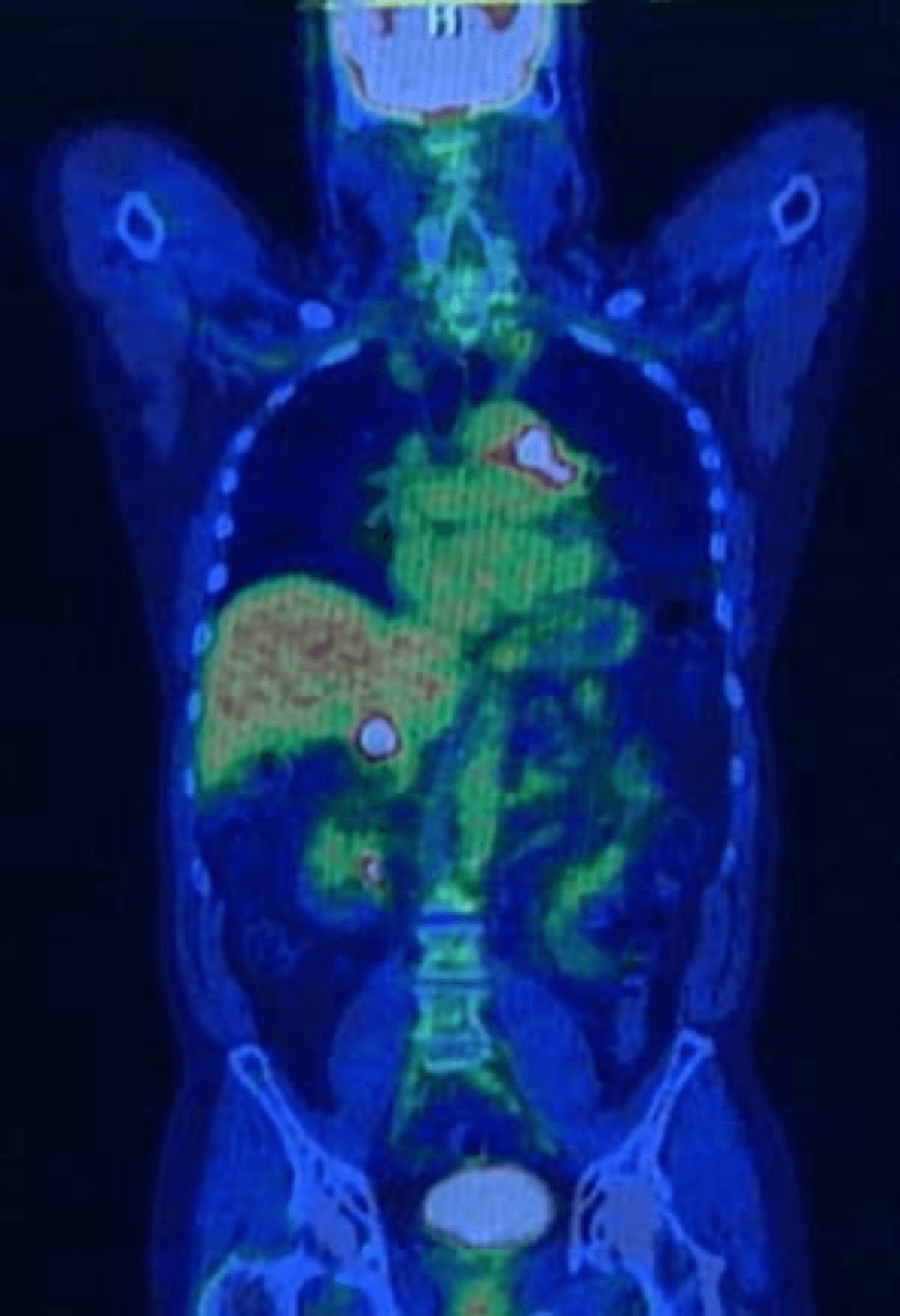
Molecular imaging uses imaging technology to identify disease by looking at the biological processes taking place at a cellular and molecular level in the body. It differs from traditional imaging in that probes known as biomarkers are used to help image particular targets or pathways. Biomarkers interact chemically with their surroundings and then alter the image according to molecular changes occurring within the area of interest.
Molecular Imaging procedures have become the standard of care in cancer treatment as well as the treatment of brain disorders, bone disorders, heart disease, kidney disease, and lung and thyroid disorders. Molecular imaging includes imaging techniques such as PET/CT and SPECT.
Depending on which type of imaging procedure is prescribed by the referring physician for the patient, the radioisotope used in Nuclear Medicine, will either be inhaled as a gas, swallowed orally in a tablet form, or injected intravenously. Once in the body the radioisotope typically accumulates in a particular area of the body or organ indicating the location of the disease, it’s size, and severity. The most commonly used radioisotope today is Flurodeoxyglucose (FDG or F18) for patients suffering with cancer. Though F-18 is the most commonly used radioisotope, other isotopes such as Ammonia (N13) and Sodium Fluoride (NaF) are also being utilized for cardiac profusion (N13) and bone scans (NaF).
The field of molecular imaging is still considered to be in its early stages and is showing a huge potential for growth as newer isotopes and scanning techniques are developed. Each of our centers have dedicated training, education, research and development areas. SAH is focused on being at the forefront of research and development in the field of molecular imaging and nuclear medicine.
How it works?
PET/CT and SPECT both require the injection of a radioactive tracer into the patient. FDG (Flurodeoxyglucose or 18F) is the most commonly used radioisotope for PET/CT becasue it accumulates in tumors and is detectable by the PET/CT scanner.
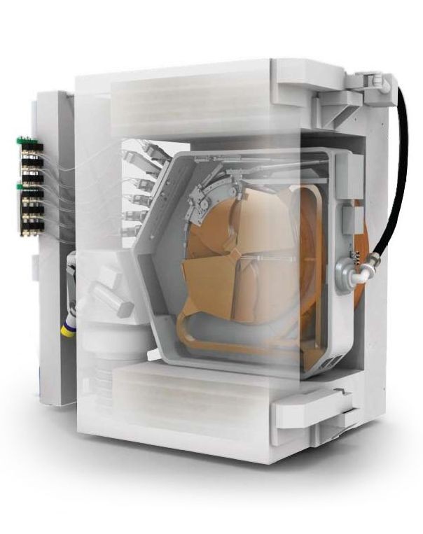
Radioactive isotopes are produced using a cyclotron and further chemical synthesis processes. The cyclotron is a particle accelerator that produces a beam of charged particles. The cyclotron consists of electrodes in a vacuum chamber that creates a magnetic field. A high-frequency alternating voltage is applied to the electrodes, which alternately attracts and repels charged particles, causing them to accelerate. The magnetic fields move the particles in a circular path and, as they gain more energy from the accelerating voltage, spiral outwards until they reach the outer edge of the chamber. The excess electrons are stripped of the ions forming positive particles such as protons or deuterons, which can be extracted as a beam. After bombardment the radioisotope is then placed in an automated synthesis unit where it undergoes a series of chemical processes to produce the final radioisotope.
SPECT is similar to PET in its use of radioactive tracer material and detection of gamma rays. In contrast with PET, however, the tracers used in SPECT emit gamma radiation that is measured directly, whereas PET tracers emit positrons that annihilate with electrons up to a few millimeters away, causing two gamma photons to be emitted in opposite directions. A PET scanner detects these emissions “coincident” in time, which provides more radiation event localization information and, thus, higher spatial resolution images than SPECT (which has about 1 cm resolution). SPECT scans, however, are significantly less expensive than PET scans, in part because they are able to use longer-lived, more easily obtained radioisotopes than PET.